Datei:Schematic diagram of the human eye.png
Beschreibung
| Beschreibung |
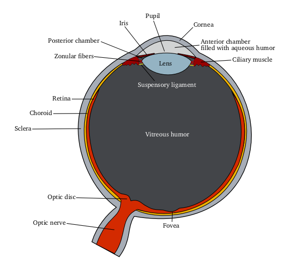
English: Schematic diagram of the human eye
|
|||||
| Quelle |
Eigenes Werk mittels: |
|||||
| Urheber | Delta G | |||||
| Andere Versionen |
[]
By languages
For translate
Anterior segment
|


Lizenz
  
|
Diese Datei ist unter der Creative-Commons-Lizenz „Namensnennung – Weitergabe unter gleichen Bedingungen 3.0 nicht portiert“ lizenziert. Vorbehaltlich der Haftungsausschlüsse. | |
| ||
| Diese Lizenzmarkierung wurde auf Grund der GFDL-Lizenzaktualisierung hinzugefügt. |

|
Es ist erlaubt, die Datei unter den Bedingungen der GNU-Lizenz für freie Dokumentation, Version 1.2 oder einer späteren Version, veröffentlicht von der Free Software Foundation, zu kopieren, zu verbreiten und/oder zu modifizieren; es gibt keine unveränderlichen Abschnitte, keinen vorderen und keinen hinteren Umschlagtext.
Der vollständige Text der Lizenz ist im Kapitel GNU-Lizenz für freie Dokumentation verfügbar. Vorbehaltlich der Haftungsausschlüsse. |
Eye Anatomy
A guide to the many parts of the human eye and how they function.
The ability to see is dependent on the actions of several structures in and around the eyeball. The graphic below lists many of the essential components of the eye's optical system.
When you look at an object, light rays are reflected from the object to the cornea, which is where the miracle begins. The light rays are bent, refracted and focused by the cornea, lens, and vitreous. The lens' job is to make sure the rays come to a sharp focus on the retina. The resulting image on the retina is upside-down. Here at the retina, the light rays are converted to electrical impulses which are then transmitted through the optic nerve, to the brain, where the image is translated and perceived in an upright position!
Think of the eye as a camera. A camera needs a lens and a film to produce an image. In the same way, the eyeball needs a lens (cornea, crystalline lens, vitreous) to refract, or focus the light and a film (retina) on which to focus the rays. If any one or more of these components is not functioning correctly, the result is a poor picture. The retina represents the film in our camera. It captures the image and sends it to the brain to be developed. The macula is the highly sensitive area of the retina. The macula is responsible for our critical focusing vision. It is the part of the retina most used. We use our macula to read or to stare intently at an object.
source: http://www.stlukeseye.com/Anatomy.asp Archivkopie in der Wayback Machine
Dateiversionen
Klicke auf einen Zeitpunkt, um diese Version zu laden.
| Version vom | Vorschaubild | Maße | Benutzer | Kommentar | |
|---|---|---|---|---|---|
| aktuell | 14:00, 5. Jun. 2006 | | 600 × 550 (54 KB) | wikimediacommons>Eliashc | optimized using optipng. |
Dateiverwendung
Die folgenden 2 Seiten verwenden diese Datei:
